
Presentation
Headache.
Patient Data
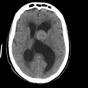

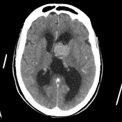
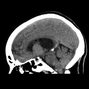
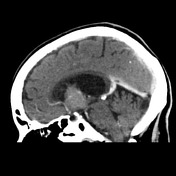
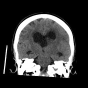
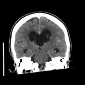

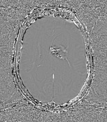
Rounded minimally enhancing lesion within the lateral ventricle near the foramen of Munro with punctate areas of calcification. Marked hydrocephalus.
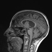
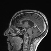
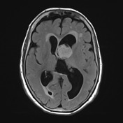

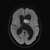
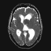
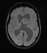
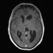
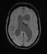
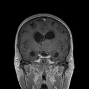

A mass within the left lateral ventricle at the junction of the frontal horn and body demonstrates predominantly peripheral high T2 signal components that suppress on FLAIR, low-grade heterogeneous enhancement and has a broad contact/attachment to the left lateral margin of the septum pellucidum. Superiorly, strands of non-enhancing tissue that demonstrates susceptibility artefact. The lesion demonstrates high DWI signal with low ADC values of approximately 650 x 10-6mm2/s. The septum pellucidum is deviated to the right by up to 12 mm.
The lesion results in moderate obstruction of the lateral ventricles with associated periventricular T2/FLAIR hyperintensities suggestive of subependymal fluid spread. No appreciable direct intra-axial extension nor further intraventricular lesion. Non-enhancing T2/FLAIR white matter hyperintensities throughout the cerebral hemispheres are most compatible with changes of chronic small vessel ischemia.
Conclusion:
Intraventricular tumor with imaging features favoring a central neurocytoma (somewhat atypical in a patient of this age) or subependymoma. Other differentials for an intraventricular mass including an ependymoma or intraventricular metastasis are felt less likely given the imaging appearances.
Case Discussion
The patient went on to have a resection.
Histology
The sections show multiple fragments of tissue which is largely replaced by a cellular neoplasm. The tumor consists of uniform formed cells which are arranged in sheets and small packets. The cells have round uniform nuclei which focally have a finely granular chromatin pattern. There is prominent clear and lightly eosinophilic granular cytoplasm. Focal areas of calcification are present within the tumor. There is also in the background a delicate vascular network present. No significant mitotic figures are identified. No vascular proliferation or necrosis is seen.
- Synaptophysin: Positive
- Chrommogranin: Negative
- CD56: Positive
- ATRX: Positive
- IDH1: Negative
- GFAP: Positive
- Ki67: <1%
Final diagnosis: central neurocytoma.


 Unable to process the form. Check for errors and try again.