
Presentation
Headache.
Patient Data


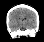
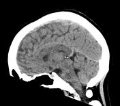
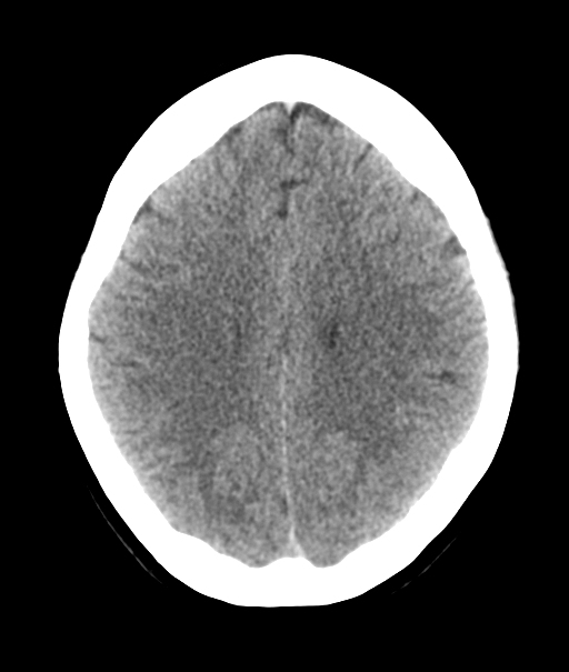
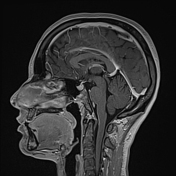
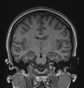
A fairly subtle mass in the region of the frontal horn and anterior body of the left lateral ventricle is present, without calcification or hydrocephalus.
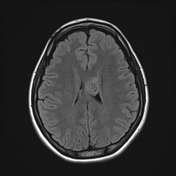
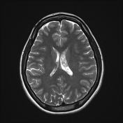
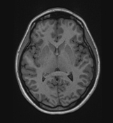
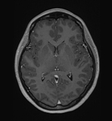
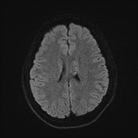
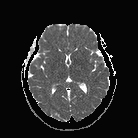
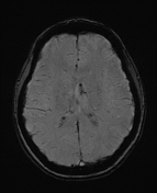

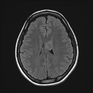
An intraventricular mass in the body of the left lateral ventricle is heterogeneously T2 hyperintense, with tiny internal cystic foci. There is a punctate focus of abnormal susceptibility in the anterior aspect of the tumor, which is favored to represent microhemorrhage rather than calcification on the basis of SWI phase images. There is minor central and peripheral punctate enhancement. The solid component of the tumor demonstrates ADC values of 1100 x 10-6 mm2/s.
The tumor has broad-based contact with the roof and lateral wall of the lateral ventricle, as well as the septum pellucidum that is minimally deviated to the right. The body of the left fornix is displaced inferomedially. No obstructive hydrocephalus. No parenchymal edema.
No other lesion is detected in the brain.
Conclusion:
This lesion most likely represents a central neurocytoma with a far less likely differential diagnosis of subependymoma, particularly in this age group.
Case Discussion
The patient went on to have a resection.
Histology
The paraffin-embedded sections show choroid plexus and tumor formed by solid sheets of a monotonous population of cells with round nuclei, granular salt-and-pepper chromatin, and indistinct cytoplasm. There is no significant nuclear atypia. Mitotic figures are not found.
Immunohistochemical stains are strong and diffusely positive for synaptophysin, NeuN and INSM1. Chromogranin, GFAP, OLIG2 and EMA are negative. IDH1 is negative and ATRX is retained.
Ki-67 = 3.5%
Final diagnosis
Central neurocytoma, WHO grade 2.


 Unable to process the form. Check for errors and try again.