Q: What are the types of Neurocytomas in the 2021 classification of tumors of the central nervous system of the World Health Organization (WHO)
show answer
A: According to the World Health Organization (WHO) 2021 classification of tumors of the central nervous system, neurocytomas are grade II brain tumors divided into two categories: - Intraventricular neurocytoma known as central neurocytoma (CNC) when located within the ventricles, which are generally slow-growing and have a low potential for malignancy; - Extraventricular neurocytoma (EVN), a rare tumor that arises anywhere in the central nervous system outside the ventricles, including the cerebral hemispheres, cerebellum, spinal cord, thalamus, pons, amygdala, pineal gland, and retina.
Q: What are the characteristics of central neurocytoma?
show answer
A: Central neurocytomas (CNs) are rare, low-grade tumors, WHO grade II (2021). They constitute approximately 0.25–0.5% of all intracranial tumors. These tumors predominantly affect adults in their second or third decade of life and appear to affect both males and females equally. Central neurocytomas grow into the ventricles, often blocking cerebrospinal fluid flow, causing hydrocephalus. Central neurocytoma origin is unknown, but it is speculated to arise from neuronal cells of the septum pellucid and the subependymal cells of the walls of the lateral ventricles.
Q: What are some of the typical clinical symptoms associated with a central neurocytoma?
show answer
A: The clinical symptoms associated with a neurocytoma can vary depending on the location and size of the tumor. The most common symptoms are due to increased intracranial pressure from hydrocephalus. Symptoms of increased intracranial pressure are headaches, vomiting, nausea, seizures, paresis, decreased consciousness, memory problems, papilledema, and visual changes. Rarely central neurocytoma may be associated with sudden death secondary to acute ventricular obstruction. Also rare is a sudden presentation due to bleeding inside the ventricles, referred to as intraventricular hemorrhage.
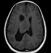
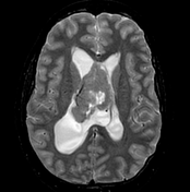
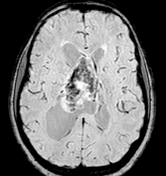
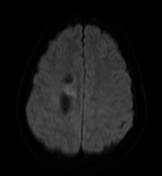
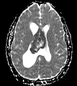
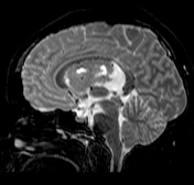
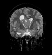
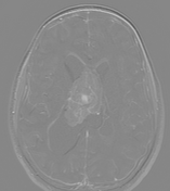
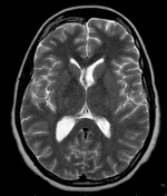
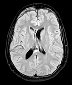
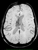
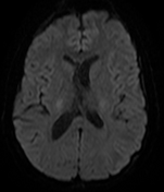
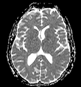
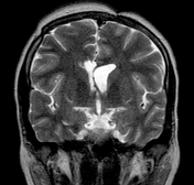
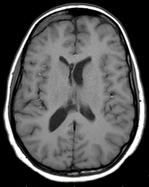
Q: What are some of the critical features of CT or MRI that can help diagnose a central neurocytoma?
show answer
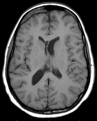
A: A central neurocytoma's cross-sectional imaging appearance is a well-circumscribed, lobulated, intraventricular mass most commonly attached to the septum pellucidum. On computed tomography (CT) scans, these tumors appear as an intraventricular dense mass, which may have punctate or coarse calcifications. On magnetic resonance imaging (MRI), central neurocytomas are isointense or hypointense on T1-weighted and isointense or hyperintense on T2-weighted and may contain small cysts, hemorrhagic areas, or calcifications. The moderate and heterogeneous enhancement pattern is typical after administering intravenous contrast on CT and MR images. Intraventricular hemorrhage may occur in patients with central neurocytoma.
Q: What are the pathologic aspects of central neurocytoma?
show answer
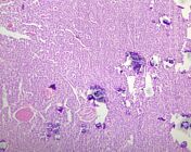
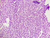
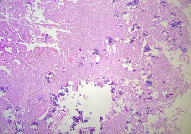
A: Central neurocytomas (CNs) appear macroscopically as a lobulated solid mass with areas of hemorrhage and may often be calcified. On histology, central neurocytomas are well-differentiated tumors composed of uniform small cells with regular round nuclei, perinuclear halos, and scant cytoplasm with stippled chromatin. Additional features include fibrillary areas closely resembling neutrophils. CN is histologically similar to oligodendroglioma and ependymoma, which can be distinguished by immunohistochemical. Atypical central neurocytoma usually presents increased mitotic activity, nuclear atypia, infiltrative margins, focal necrosis, vascular proliferation, and Ki-67 index > 2%,
Q: What are some of the immunohistochemical markers?
show answer
A: Some immunohistochemical markers help diagnose CNs. These tumors show positive immunoreactivity for synaptophysin, neuron-specific nuclear protein (Neu-N), and neuron-specific enolase (NSE). Immunohistochemically, it is usually negative for epithelial membrane antigen (EMA), glial fibrillary acidic protein (GFAP), and vimentin. The MIB-1 LI is a prognostic tool to determine tumor relapse.
Q: What is the goal of treatment for a central neurocytoma?
show answer
A: The treatment approach for a central neurocytoma depends on the tumor's size, location, and grade. Gross total resection (GTR) is often the primary treatment option for CNs. Patients receiving subtotal resection (STR) have an increased risk for recurrence, and clinicians may also treat them with adjuvant radiotherapy or radiosurgery and, occasionally, chemotherapy. However, the efficacy of these treatments is less well-established.
Q: What is the differential diagnosis of central neurocytoma?
show answer
A: Ependymoma, subependymoma, choroid plexus papilloma, intraventricular meningioma, and intraventricular metastasis are differential diagnoses for central neurocytoma.


 Unable to process the form. Check for errors and try again.