
Presentation
Ataxia.
Patient Data


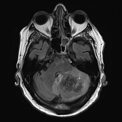
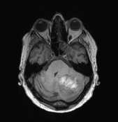
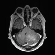
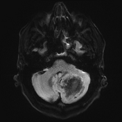
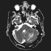
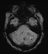
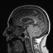
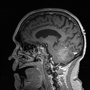
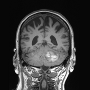
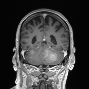
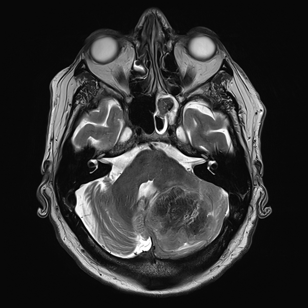
Multiple cerebellar lesions are identified, the largest in the left hemisphere (50 mm in diameter) and three smaller ones in the right hemisphere. The lesions have intrinsic high T1, only slight contrast enhancement, and profoundly low T2 signal with minimal loss of signal on SWI.
The fourth ventricle is distorted. Ventricles are mildly prominent without overt obstructive hydrocephalus and fairly pronounced cerebral volume loss. No visible supratentorial lesions.
Conclusion
Imaging appearances are those of metastases. The likely primarys include melanoma and adenocarcinoma (likely mucinous).
Case Discussion
This patient had a known diagnosis of esophageal carcinoma. They went on to have a resection.
Histology
Sections show cerebellar tissue infiltrated by complex glands and sheets of malignant cells, and abundant associated necrosis. Cells have large, variable nuclei, prominent nucleoli, and mitoses are frequent.
The tumor is positive for CK7 and CK19, with patchy positive staining for CK20 and CDX-2. It is negative for TTF-1, GATA-3, PAX-8, and NKX3.1. HER-2 shows diffuse strong and complete membranous staining (3+).
Final diagnosis
Metastatic adenocarcinoma consistent with an upper gastrointestinal primary.
Discussion
This case illustrates the typical profoundly low T2 signal seen in adenocarcinoma metastases. This is usually attributed to the presence of mucin, although there is some debate as to whether this is the case and if so, what the mechanism for T2 shortening is 1,2.


 Unable to process the form. Check for errors and try again.