Presentation
Follow-up brain MRI after brain tumor resection 3 years ago.
Patient Data
Age: 35 years
Gender: Female
From the case:
Cerebellar pilocytic astrocytoma - recurrence
![](https://prod-images-static.radiopaedia.org/images/56236221/011_thumb.jpeg)
![This study is a stack](/packs/stack-YQKLCKBI.gif)
![](https://prod-images-static.radiopaedia.org/images/56236242/012_thumb.jpeg)
![This study is a stack](/packs/stack-YQKLCKBI.gif)
![](https://prod-images-static.radiopaedia.org/images/56236282/010_thumb.jpeg)
![This study is a stack](/packs/stack-YQKLCKBI.gif)
![](https://prod-images-static.radiopaedia.org/images/56236300/010_thumb.jpeg)
![This study is a stack](/packs/stack-YQKLCKBI.gif)
![](https://prod-images-static.radiopaedia.org/images/56236321/013_thumb.jpeg)
![This study is a stack](/packs/stack-YQKLCKBI.gif)
![](https://prod-images-static.radiopaedia.org/images/56236357/025_thumb.jpeg)
![This study is a stack](/packs/stack-YQKLCKBI.gif)
![](https://prod-images-static.radiopaedia.org/images/56236390/010_thumb.jpeg)
![This study is a stack](/packs/stack-YQKLCKBI.gif)
![](https://prod-images-static.radiopaedia.org/images/56236410/012_thumb.jpeg)
![This study is a stack](/packs/stack-YQKLCKBI.gif)
![](https://prod-images-static.radiopaedia.org/images/56236432/012_thumb.jpeg)
![This study is a stack](/packs/stack-YQKLCKBI.gif)
Download
Info
![](https://prod-images-static.radiopaedia.org/images/56236221/011_big_gallery.jpeg)
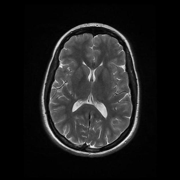
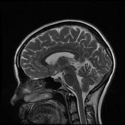
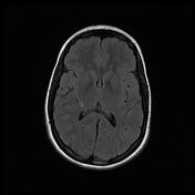
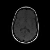
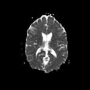
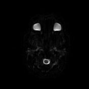
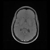
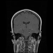
Left cerebellar hemisphere cystic lesion with a solid mural nodule that displays moderate contrast enhancement
Post-treatment changes at the left-sided posterior fossa.
Case Discussion
Gradual regrowth of the known brain tumor (histopathologically proven case of pilocytic astrocytoma) three years after surgical resection has been shown on follow-up brain MRI.