Presentation
Headaches.
Patient Data
Age: 45 years
Gender: Female
From the case:
Cerebral cavernous malformation


Download
Info
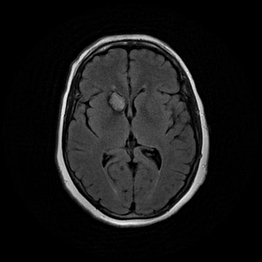
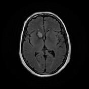
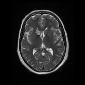
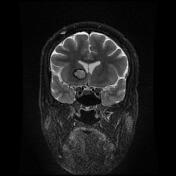
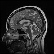
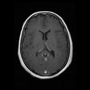
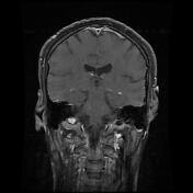
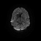
There is a 15 x 20 mm well-defined heterogenous signal mass lesion in the right caudate nucleus and anterior limb of the internal capsule, with a dark hemosiderin ring and popcorn appearance typical for cavernous angioma.
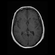
After contrast injection, the mass shows internal enhancement.
Few high signal foci in T2 and flair sequences at subcortical white matter of both cerebral hemispheres depict microvascular ischemic events.
Case Discussion
MRI is the modality of choice, demonstrating a characteristic “popcorn” or "berry" appearance with a rim of signal loss due to hemosiderin that confirmed the diagnosis of a cavernoma.


 Unable to process the form. Check for errors and try again.