
Presentation
Headache.
Patient Data
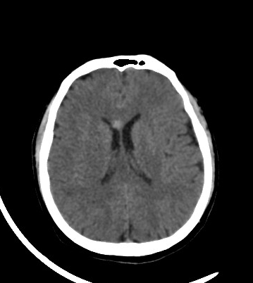
Hyperattenuating relatively well-defined oval lesion seen in the genu of corpus callosum right to the midline, measuring about 0.8 x 0.9 cm and without evidence of surrounding oedema.
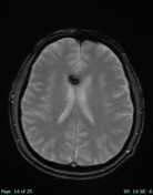

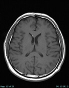
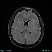
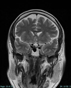
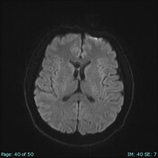
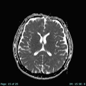
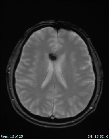
There is a well a circumscribed lesion with mixed signal on T2 and blooming artifact on gradient echo (SWI), without surrounding oedema noted in the genu of corpus callosum right to the midline, measuring about 1.2 x 1.2 cm. No evidence of restricted diffusion of the lesion. Findings are suggesting cavernoma.
Linear blooming artefact are seen in the left cerebellar hemisphere suggesting developmental venous anomaly.
Case Discussion
Cerebral cavernous venous malformations account for 5-15% of the vascular malformation of the central venous system.
Magnetic resonance imaging is an important modality to diagnose and evaluate cavernous malformations, especially, susceptibility-weighted imaging which is considered highly sensitive sequence for cavernous malformations.
There are 4 types of cerebral cavernous venous malformations based on Zabramski classification. This case represents type II, which is the most common type.


 Unable to process the form. Check for errors and try again.