
Presentation
Headache and fits for a few months.
Patient Data
Age: 55 years
Gender: Female
From the case:
Cerebral cavernous venous malformation

Show annotations
Download
Info

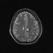
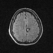
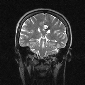
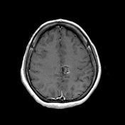
A popcorn-like, altered signal intensity lesion involving the left medial frontoparietal regions adjacent to the flax cerebri is noted. The returning signals are hyperintense T2 and FLAIR, and the post-contrast images show no significant enhancement. No perilesional edema is seen. The area is surrounded by a T2 and FLAIR hypointense ring, suggesting a rim of hemosiderin.
Case Discussion
MRI features are of a vascular lesion involving the left frontoparietal region, most likely suggestive of cavernous hemangioma (cavernoma).


 Unable to process the form. Check for errors and try again.