
Presentation
Weakness of the lower limbs.
Patient Data
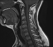

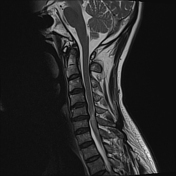
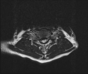
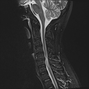
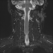
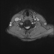
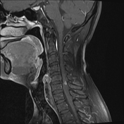
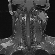
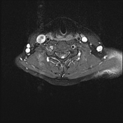
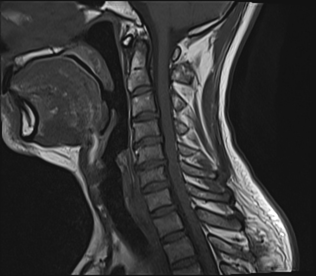
MRI of the cervical spine with contrast enhancement shows a central protrusion of a herniated disc at C5/6 and osteophyte formation resulting in severe spinal canal stenosis (grade 3 according to Kang), severe bilateral foraminal stenosis, and deformation and edema of the spinal cord at the same level. The edema extends to the spinal cord at the C6/7 level, with hyperintense signals on T2W and STIR sequences. After contrast administration, there is strong nodular enhancement at the C5/6 spinal cord level. Reduced vertebral body height, disc desiccation, and facet joint degeneration are also observed.
Case Discussion
The MRI findings are consistent with cervical compressive myelopathy caused by disc herniation combined with degenerative changes in the cervical spine.
Note that a small percentage of these lesions show contrast enhancement after injection. The patient subsequently underwent surgery for herniated disc removal and spinal cord decompression.


 Unable to process the form. Check for errors and try again.