
Presentation
Painless swelling of the right parietal region with recent neck pain.
Patient Data


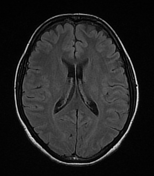
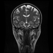
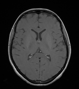
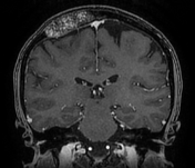
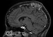

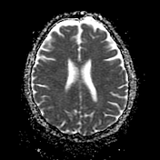
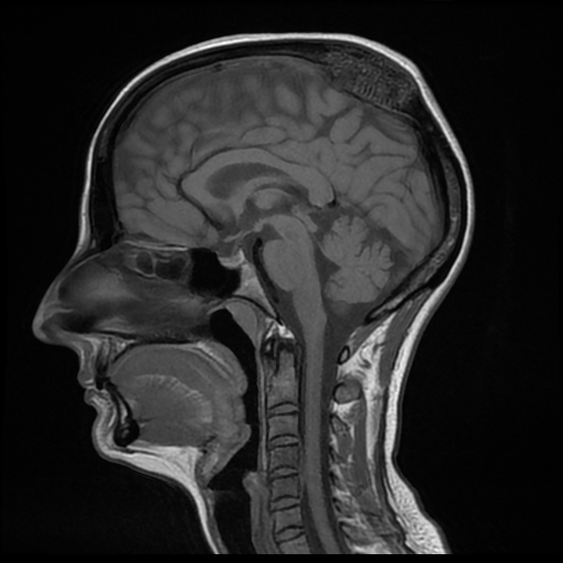
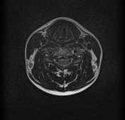
There is an expansile coarse trabeculated intraosseous lesion of the right parietal bone with thinning of the internal and external skull tables. It appears non-homogeneously, hypo-to-isointense on T1WI, hyperintense on FLAIR and T2WI with inhomogeneous enhancement following IV contrast. A mass effect is noted on the adjacent parietal lobe as well as the superior sagittal sinus which remains patent. No intra-axial extension seen.
In the cervical region on the sagittal T1 sequence, there is an abnormal appearance at the C4-C5 level.
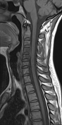
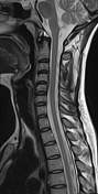
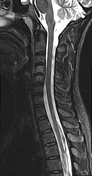
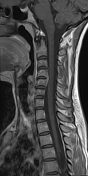
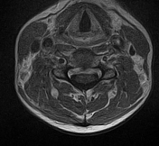

The patient was called and an MRI of the cervical spine was performed 5 days later. It shows a subtle narrowing of C4-C5 disc space of intermediate signal intensity on T1WI with area high signal intensity on T2WI and STIR. The C4 and C5 vertebral bodies appear of low signal intensity on T1WI, high signal intensity on T2WI with enhancement on post-contrast sequences (bone marrow edema). No evidence of destruction of the neighboring endplates. Note a fusiform soft tissue mass in the anterior epidural space at the C4-C5 level of intermediate signal intensity on both T1WI and T2WI with homogeneous enhancement following IV contrast (epiduritis).
Case Discussion
MRI features of a C4-C5 spondylodiscitis in a patient with skull vault hemangioma
In such case, it is important that the radiologist must take in consideration the clinical data during the visualization and interpretation of any radiological examination.


 Unable to process the form. Check for errors and try again.