
Presentation
Knee pain and swelling
Patient Data


Expansile lesion of the proximal tibia with sharp zones of transition and expansion. It reaches the articular surface without breach of the cortex here or elsewhere. No matrix calcification or soft tissue component.


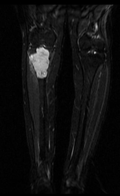
MRI demonstrates the lesion to have a very high T2 signal, low T1 signal with diffuse somewhat heterogeneous contrast enhancement. There is no surrounding edema (neither in the bone nor in the adjacent soft tissues).
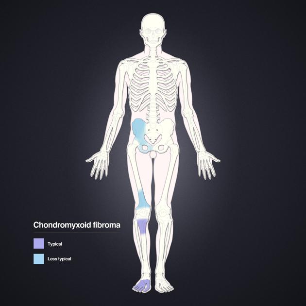
Distribution of chondromyxoid fibromas
Distribution of chondromyxoid fibromas. Layout and distribution: Frank Gaillard 2012, Line drawing of skeleton: Patrick Lynch 2006, Creative Common NC-SA-BY

Bone scan demonstrates thin peripheral increased activity. It is an isolated abnormality.
Case Discussion
This is an advanced case of a benign chondromyxoid fibroma. Smaller lesions are typically located in the cortex of long bones. Tibia and foot are most commonly affected. The lesion is never purely epiphyseal, but may extend into the subchondral bone.


 Unable to process the form. Check for errors and try again.