
Presentation
Diplopia.
Patient Data


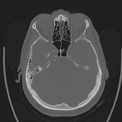
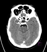

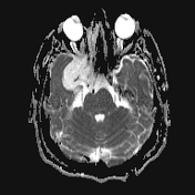
A mass in the right middle cranial fossa is heterogeneous in density, with areas of calcification admixed to areas of density similar to brain parenchyma and other areas of lower attenuation. It has a well-defined thin sclerotic margin medially; the remodeled lateral wall of the sphenoid sinus. Laterally, it displaces the temporal lobe.
There is also destruction of the right carotid canal with extension into the clivus and to the posterior margin of foramen ovale. The right hypoglossal canal is breached superiorly.
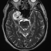
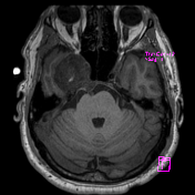
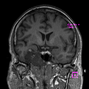
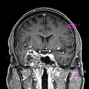
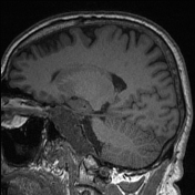
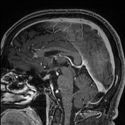
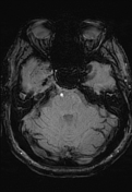

A well-defined T2 hyperintense lobulated mass centered at the right clivus, crossing the midline by 4 mm, is heterogeneous but predominantly T1 hypointense, brightly T2 hyperintense, with loculated/bubbly internal enhancement.
There is a breach of bony cortex posteriorly with a projection into the prepontine cistern beneath the dura but lying in close proximity to the basilar artery.
The right cavernous carotid artery is displaced, but its course lies predominantly inferomedial. Normal cavernous carotid flow void is preserved, although mild narrowing is present. The right cavernous sinus and Meckel's cave are obliterated.
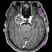
The tumor lies close to the right optic canal, but the optic canal is not compromised. Inferiorly, a tongue of tissue has protruded into the right longus capitis muscles but is 7mm from the right nasopharyngeal mucosa.
Right cochlear and vestibular apparatuses, middle ear and mastoid uninvolved. Right jugular foramen also appears uninvolved.
Conclusion:
Right central skull base tumor most likely to represent chondrosarcoma or, less likely, (eccentric) chordoma.
Case Discussion
The patient went on to have a resection.
Histology
Paraffin sections show a tumor of varying hypercellularity composed largely of circumscribed lobules of chondroid material in which tumor cells are haphazardly arranged in lacunae. Many lacunae contain multiple tumor cells. Tumor cells show mild nuclear atypia. An occasional mitotic figure is identified. Other areas of tumor are composed of single and small aggregates of tumor cells dispersed in a mucinous/myxoid stromal matrix. No areas of necrosis are identified. Lobules of chondroid tumor tissue are seen to permeate between trabeculae of native bone. Moderate bone remodeling is seen.
Final diagnosis
Chondrosarcoma - Grade II
Discussion
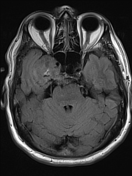
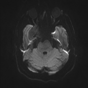
This is fairly typical appearance of a chondrosarcoma, demonstrating internal rings and arcs ossification and very high T2 signal, located lateral to the midline, centered on the petroclival synchondrosis.


 Unable to process the form. Check for errors and try again.