
Patient Data
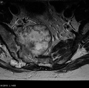
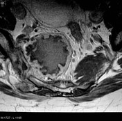

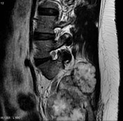
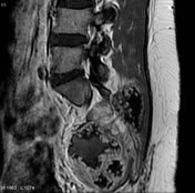
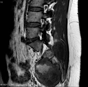

Destructive sacral mass with a large extraosseous component in the sciatic notch and pelvis.
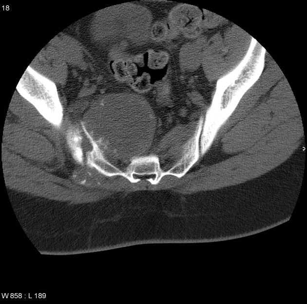
Solitary CT axial image through the sacrum demonstrates a mass arising from the right sacral ala with a large component in the pelvis.
Case Discussion
The patient went on to have a resection preceded by preoperative core-biopsy.
Histology
Preoperative core biopsy: the lesion appears well-sampled and is comprised of disorganized cartilage, with some areas of myxoid change. Chondrocyte necrosis is present, dissociated from calcification.
Resection specimen
Complex resection of right hemisacrum, sacroiliac joint, medial ilium, proximal sciatic nerve, and associated soft tissues. Tumor (9cm) is based in the sacroiliac joint but invades the adjacent portions of sacrum and ilium, as well as skeletal muscle and soft tissues anterior and posterior to the SI joint. All resection margins negative. No vascular invasion.
Final diagnosis: conventional chondrosarcoma - grade 2


 Unable to process the form. Check for errors and try again.