

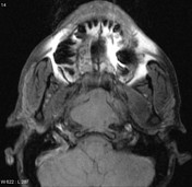
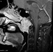
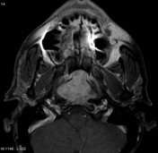


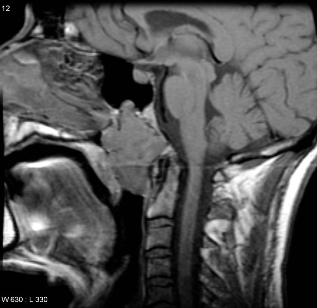
MRI through the base of skull demonstrates a large midline mass centered on the clivus, bulging into the nasopharynx. It is intermediate in T1 signal and markedly hyperintense in T2 signal and demonstrates homogeneous enhancement.
Case Discussion
Features are characteristic of a chordoma which was subsequently confirmed on biopsy.
Histology
Microscopic Description: Sections show a lobulated tumor with cells arranged in cords and sheets, present in a predominantly myxoid and focally sclerotic stroma. Some tumor cells exhibit abundant vacuolated cytoplasm (physaliferous appearance). Infiltration into surrounding fibromuscular tissue is observed. Areas of extensive fibrosis are associated with mixed chronic inflammatory infiltrates and focal hemosiderin deposition. Immunohistochemical studies show the neoplastic cells to be immunoreactive for keratin, EMA, and focally for S100. The reactive inflammatory infiltrates display immunoreactivity for CD20 and CD3.
FINAL DIAGNOSIS: chordoma


 Unable to process the form. Check for errors and try again.