
Patient Data


A subtle soft tissue density nodule is seen at the level of L3/4 without bony destruction or remodeling.


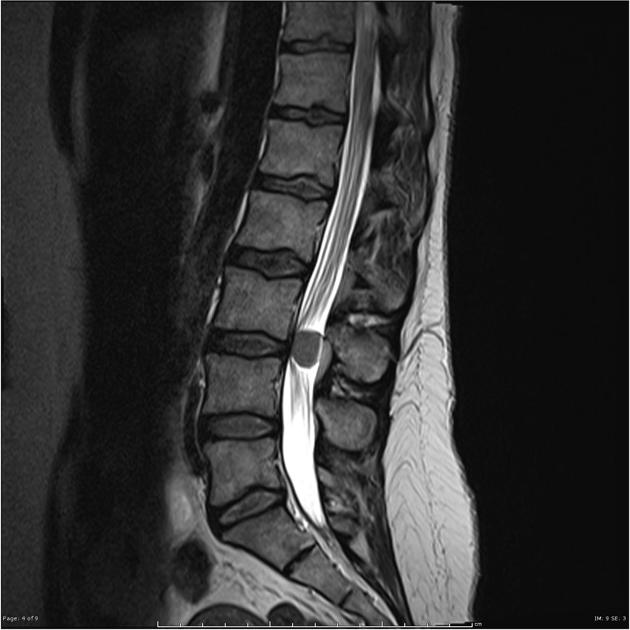
Vividly enhancing intradural mass is present at the level of L3/4, below the tip of the conus. It displaces the majority of the cauda equina to the left, and does not clearly arise from never roots.
Differential includes: meningioma or a neurogenic tumor (e.g. schwannoma) or a myxopapillary ependymoma. Its signal intensity and apparent thecal base favors a meningioma.


MACROSCOPIC APPEARANCE:
"L3/4 spinal tumor": An ovoid piece of rubbery tan and pink tissue 20x14x11mm.
MICROSCOPIC DESCRIPTION: The sections show a moderately cellular tumor. The tumor forms sheets and nests in a fibrous background. No whorls are seen. The tumor cells have round hyperchromatic nuclei, inconspicuous nucleoli and moderate amounts of clear cytoplasm. There is no nuclear pleomorphism. Mitoses are inconspicuous. There is no necrosis. The tumor cells are focally PAS positive. They are EMA and progesterone receptor positive. The Ki-67 index is about 8%. They are CAM5.2 and CD10 negative. The features are those of clear cell meningioma, which is classified as WHO grade II. DIAGNOSIS: 1-3. L3/4 spinal tumor: Clear cell meningioma (WHO Grade II).


 Unable to process the form. Check for errors and try again.