
Presentation
MVA trauma.
Patient Data
Age: 20 year-old
Gender: Male

Download
Info
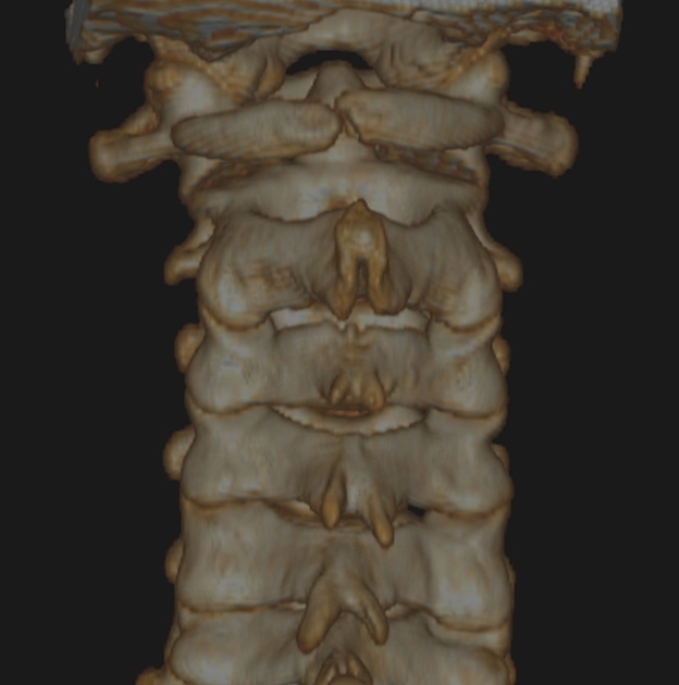
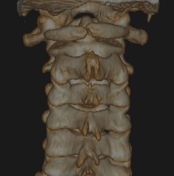
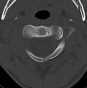
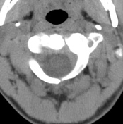
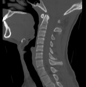
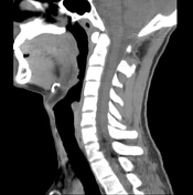
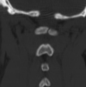
Median cleft in the posterior arch of the Atlas (C1) with regular borders lined by cortical bone. Remainder vertebral bodies, discs, spinal canal, and soft tissue are normal.
Case Discussion
This case illustrates a congenital anomaly of the posterior atlas (C1) arch characterized by its incomplete posterior fusion leading to a cleft. Like in this case, the main significance of this finding is when a fracture must be excluded. The absence of other associated abnormalities, the atypical pattern considered the trauma kinetic and the cortical lining the edges of the cleft help to make this differentiation.


 Unable to process the form. Check for errors and try again.