
Presentation
Middle aged male with headaches.
Patient Data


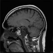

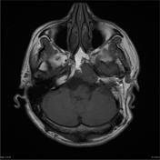
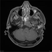
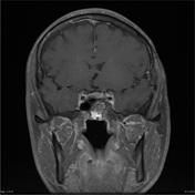
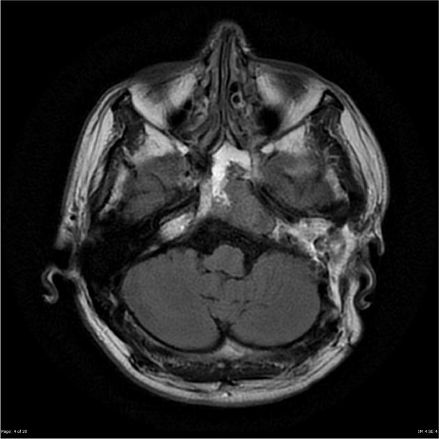
Soft tissue mass in the left side of the clivus extending into the tip of the left petrous bone and the left cerebello- pontine angle. The lesion has a markedly prolonged T2, producing marked hyperintensity within it on T2 weighted scans. A separated section of the mass is in the left side of the pons and lower mid brain. This part of the lesion abuts the basilar artery anteriorly. Anteriorly the mass extends into the sphenoidal sinus and across the midline to the right. Contrast enhancement is relatively limited and heterogeneous.
Conclusion Appearance in keeping with large chordoma.
Case Discussion
Pathology report:
The section shows fragments of a moderately hypercellular tumor. Tumor cells have small round and oval hyperchromatic nuclei and a large amount of cytoplasm with a physaliferous appearance. Much intra and extracellular mucin is noted. Tumor cells are arranged in lobules delimited by collagenous septa. A very occasional mitotic figure is identified. There is no evidence of necrosis. The features are of chordoma.
DIAGNOSIS: Clival tumor: Chordoma.


 Unable to process the form. Check for errors and try again.