
Presentation
Right elbow dislocation after falling. Closed reduction performed immediately after, with 'below elbow' back slab applied.
Patient Data

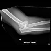

There is an acute dislocation of the right elbow joint with the distal humerus displaced medially. Tiny densities seen adjacent to the distal humerus may represent tiny avulsion/impaction fracture fragments. Significant soft tissue swelling surrounding the elbow is also noted.
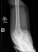

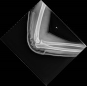
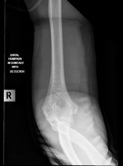
Post closed reduction of right elbow dislocation on back slab with satisfactory alignment is seen.
Case Discussion
In this case of an elbow dislocation, a closed reduction was performed to manipulate the elbow joint into its anatomical position.
Of note in this case is the radiographer's use of the elbow (acute flexion AP) views after closed reduction. This was attempted as the patient was unable to fully extend their elbow joint due to their hardened back slab. This technique results in two images being acquired, to allow true anatomical demonstration of the distal humerus and proximal forearm.
It is also imperative for all performing radiographers to note that the presence of the back slab means a higher exposure factor is required to visualize bony trabeculae and cortical outlines better.


 Unable to process the form. Check for errors and try again.