
Note: This case has been tagged as "legacy" as it no longer meets image preparation and/or other case publication guidelines.

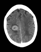
A high-attenuation mass with with a small area of central low density and surrounding minor edema demonstrates intense enhancement on post-contrast.
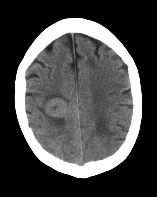
There is the suggestion of leptomeningeal enhancement elsewhere, most notably in the right parietal region; however, this is difficult to confirm on a single image.

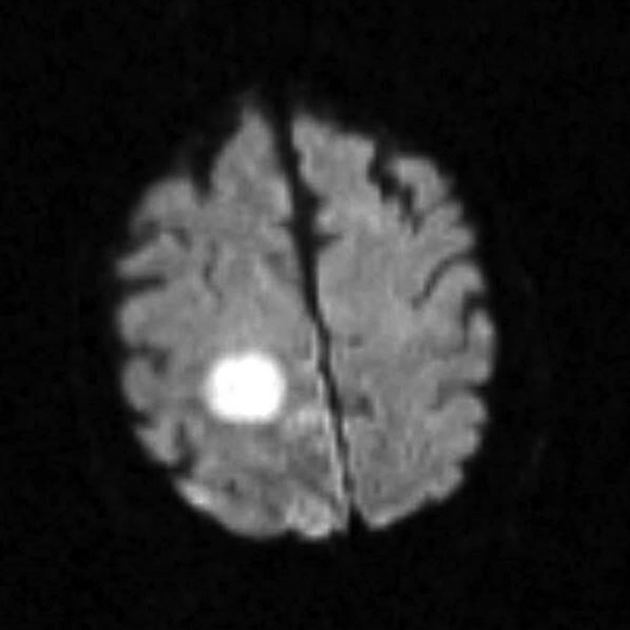
The lesion seen on CT demonstrates marked diffusion restriction (high DWI signal, and low ADC values, much lower than adjacent brain).
Case Discussion
This patient had known systemic lymphoma, and thus, this represents secondary CNS lymphoma.
The typical appearance in patients with normal immunity is a solitary enhancing mass. This is typically of high density on non-contrast CT and demonstrates restriction on DWI.
Image courtesy of R. Larsen and A. Coulthard. Royal Brisbane and Women’s Hospital, Brisbane, Australia.


 Unable to process the form. Check for errors and try again.