
Presentation
A VP shunt was inserted one year ago for obstructive hydrocephalus.
Patient Data
Age: 65 years
Gender: Female
From the case:
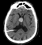
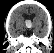
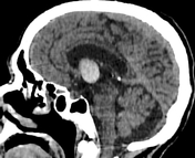
Colloid cyst of the 3rd ventricle

Download
Info

A well-defined spontaneously hyperdense ovoid lesion is seen at the roof of the 3rd ventricle measuring (2.6 x 2 x 1.8 cm), obstructing the foramen of Monro bilaterally with moderate dilatation of the lateral ventricles (obstructive hydrocephalus).
The tip of the ventriculoperitoneal (VP) shunt is well-visualised at the occipital horn of the right lateral ventricle.
Case Discussion
MRI features of a colloid cyst of the 3rd ventricle causing an obstructive hydrocephalus.


 Unable to process the form. Check for errors and try again.