
Presentation
Bilateral lower back and right groin pain. Compression L1?
Patient Data
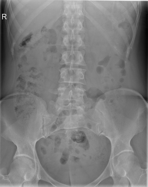
Vertebral body S1 is lumbarized. This is complete on the left side and partial on the right side, where there is an articulation between S1 and the remainder of the sacrum. Also note that there are five vertebrae above it that have no ribs, indicating that they are of lumbar nature.


The vertebra appearing as L5 is actually a hemilumbalired S1 vertebra.
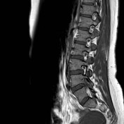
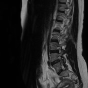
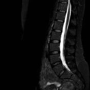
There is an empty neuroforamen on the level of S1-S2 on the right side. The S1 nerve root exits one level higher together with the L5 nerve root as can be seen both on sagittal and transverse images.
Case Discussion
Beware of conjoined nerve roots and variant anatomy of the lumbosacral spine. In this case the x-ray nicely shows that S1 is lumbarized completely on the left side and partially on the right side. This can trick you into miscounting the exiting nerve roots.


 Unable to process the form. Check for errors and try again.