
Presentation
Presented with an episode of pulmonary edema.
Patient Data
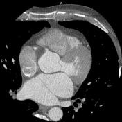

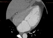
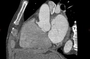
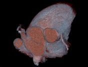

Retrospectively gated cardiac CT clearly demonstrates fibromuscular membrane dividing left atrium into a proximal chamber, containing pulmonary venous confluence, and a distal "true" left atrium, which contains the left atrial appendage. Membrane has large fenestration in its lower portion allowing decompression of pulmonary veins.
Case Discussion
Cor triatriatum sinister is a rare congenital cardiac anomaly in which the pulmonary venous confluence is separated from "true" left atrium by fibromuscular septum. Cor triatriatum can persist unrecognized into adult life if large opening or several fenestrations are present. Late onset of symptoms is usually caused by increased pulmonary pressure, development of atrial arrhythmias or mitral valve abnormalities. Nonobstructive cor triatriatum may be an incidental finding.


 Unable to process the form. Check for errors and try again.