
Presentation
Previous CT confirmed hemorrhagic stroke. Developed another stroke thereafter.
Patient Data
Age: 60 years
Gender: Female
Download
Info
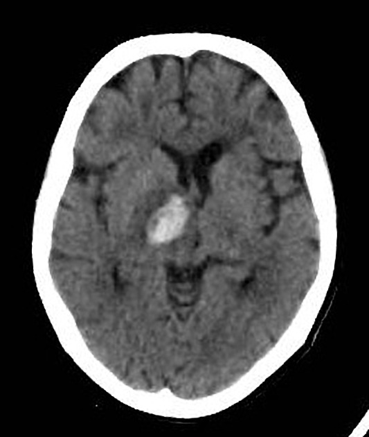
This is the first CT for the patient which shows a focus of ICH centered at the right basal ganglia, with extension into the ventricular system. Also, areas of hypodensity of different temporality at the right posterior parietal and the left deep white matter.
Download
Info
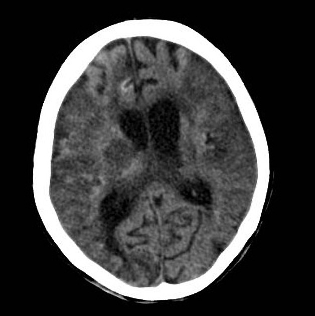
Areas of hyperdensity that follow the cerebral cortex layer at both MCA territories (gyriform), more conspicuous at the right insula, with diffuse parenchymal hypodensity indicating bilateral MCA infarcts.


 Unable to process the form. Check for errors and try again.