
Presentation
Patient presents with sudden onset visual loss left eye. O/E left retinal arterial occlusion, ? source of embolus
Patient Data

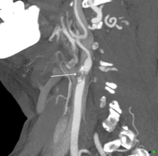
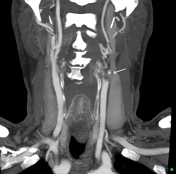
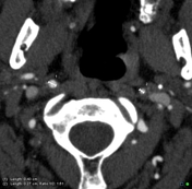

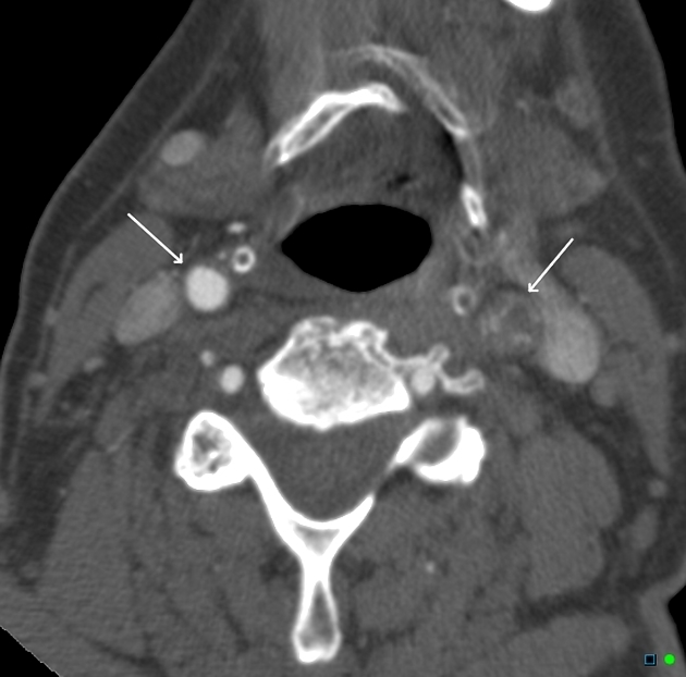
High-grade stenosis due to a calcified plaque at the origin of the left internal carotid artery. Note the internal carotid above this lesion remains patent but of reduced caliber (4.3 v 2.7 mm) and shows reduced enhancement in the terminal L ICA = 287 HU compared with R ICS = 437 HU.
Case Discussion
One indication that I use to determine the severity of internal carotid stenosis is the differential enhancement in the distal carotid lumens (beyond the stenosis). In this case, there is a 34% reduction, in contrast, density on the left c/w the right. The internal carotid artery beyond the stenosis is also of reduced caliber compared with the right due to the reduced blood flow.


 Unable to process the form. Check for errors and try again.