
Presentation
Delayed mental milestones. Normal head size. Recent onset of abnormal behaviors.
Patient Data
Age: 20 years
Gender: Male
From the case:
Cystic leukoencephalopathy without megalencephaly


Download
Info
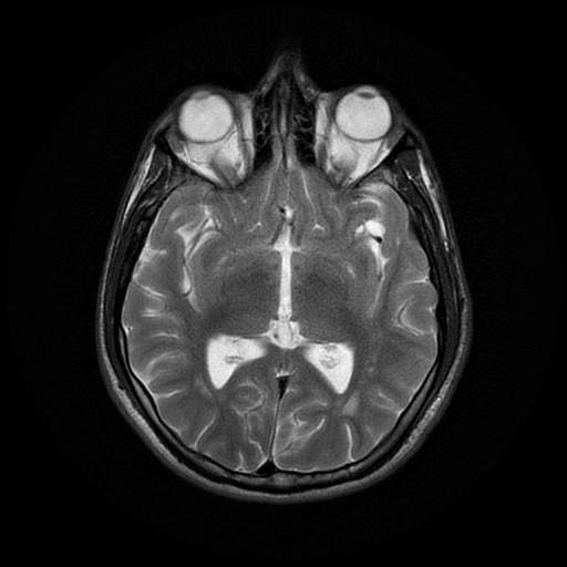
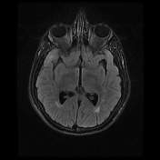
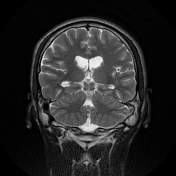
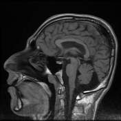
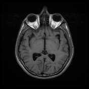
Multiple areas of altered white matter signal (low in T1 and high in T2 and FLAIR images) is demonstrated in the bilateral corona radiata semiovale regions and subcortical region of both temporal lobes accompanied with bilateral symmetrical temporal lobe subcortical cysts.
Case Discussion
A young patient with a series of MRIs since the age of 8 years, all of them exhibit the same findings of abnormal white matter signal as well as temporal subcortical cysts. all of the findings are in keeping with Cystic leukoencephalopathy without megalencephaly


 Unable to process the form. Check for errors and try again.