
Presentation
Occipital hemorrhage. Small AVM resected 7 days earlier.
Patient Data
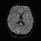

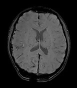
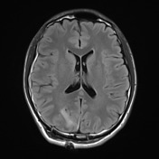
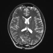


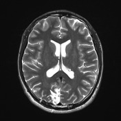
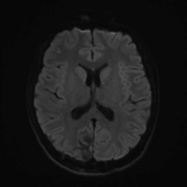
Expected postoperative changes in the right occipital lobe are seen consistent with the evacuation of a hematoma and resection of a small AVM 7 days previously. In the splenium of the corpus callosum, exactly in the midline, is an ovoid region of abnormal restricted diffusion (ADC values <500 x 10-6 mm2/s). A similar, less well-defined lesion is seen in the right corona radiata. These demonstrate reduced T1 signal, no enhancement and increased T2 signal. They may both represent ischemic foci, possibly related to surgery or catheter angiography. The splenial lesion may also represent a cytotoxic lesion of the corpus callosum (CLOCC).


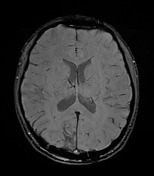
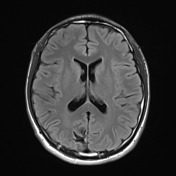
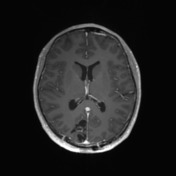
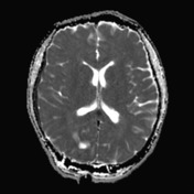
Repeat MRI 6 weeks later demonstrates the continued evolution of postoperative changes. The corona radiata lesion persists as an area of gliosis. The callosal lesion, on the other hand, has completely resolved with no lingering signal or volume change.
Case Discussion
This case illustrates typical appearances of a cytotoxic lesion of the corpus callosum (CLOCC) also known by a variety of other names including: transient lesions of the splenium of the corpus callosum, mild encephalitis/encephalopathy with a reversible isolated SCC lesion (MERS), reversible splenial lesions and reversible splenial lesion syndrome (RESLES).
Regardless of the name, these represent non-ischemic lesions seen in a variety of causes (including intracranial hemorrhage) that completely resolve.


 Unable to process the form. Check for errors and try again.