
Presentation
Recurrent wrist and hand pain for the past year. CT scan of the hand and wrist is requested for evaluation of osseous structures.
Patient Data
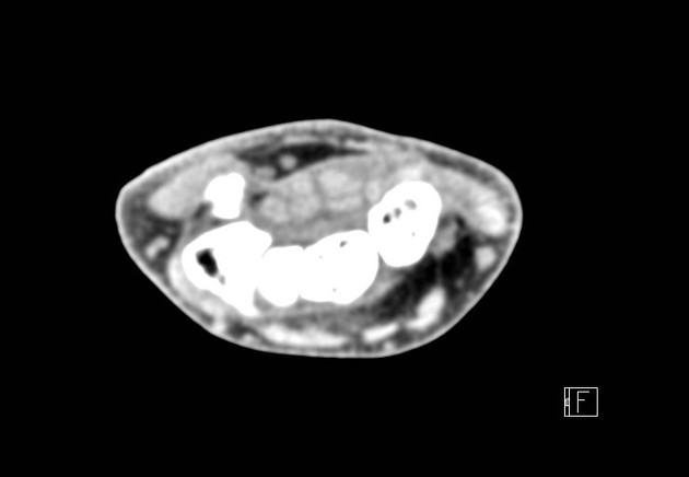
Non-contrast CT scan of the wrist and the hand demonstrates focal infiltration and thickening of the first extensor tendon compartment of the wrist. No osseous or articular abnormality is detected.
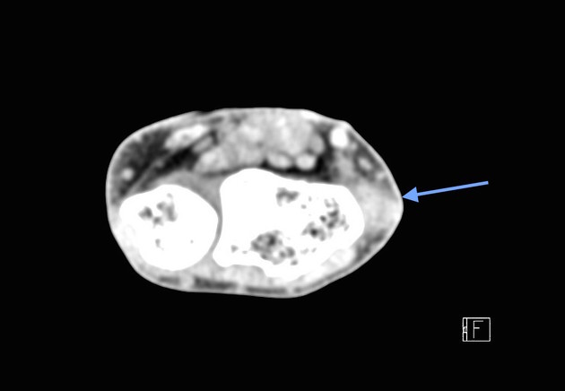
Non-contrast CT scan of the wrist and the hand demonstrates focal infiltration and thickening of the first extensor tendon compartment of the wrist (blue arrow).
Case Discussion
This case demonstrates a rare example of de Quervain tenosynovitis diagnosed on a CT scan. US and MRI are the main imaging modalities used to diagnose this entity, whereas plain film can help exclude other possibilities such as fracture, osteomyelitis or trapezio-metacarpal osteoarthritis. CT has limited interest in the workup due to its inferior soft-tissue contrast capabilities compared to US and MRI as well as radiation concerns.


 Unable to process the form. Check for errors and try again.