Presentation
Wrist pain in the radial side for seven months.
Patient Data
![](https://prod-images-static.radiopaedia.org/images/60509768/789460170d1a114afe216b3a0ac33f010b2ba37fca5b00f5181b1d14821ca0be_thumb.jpeg)
![This study is a stack](/packs/stack-YQKLCKBI.gif)
![](https://prod-images-static.radiopaedia.org/images/60509782/a79218c980859a2e7c90651e248ff9df198047569aebf53001189fbc65a8472e_thumb.jpeg)
![This study is a stack](/packs/stack-YQKLCKBI.gif)
![](https://prod-images-static.radiopaedia.org/images/60509700/250a7567a55bade4f03838a066b9886f5e5bd2783f3a1afaa238dd84cadb7c20_thumb.jpeg)
![This study is a stack](/packs/stack-YQKLCKBI.gif)
![](https://prod-images-static.radiopaedia.org/images/60509604/771abfbcc2dacb68aa6fb1849f646cdec02289059d9a8a4d5e7075c82a58237b_thumb.jpeg)
![This study is a stack](/packs/stack-YQKLCKBI.gif)
![](https://prod-images-static.radiopaedia.org/images/60509725/e8dcc5106a1299f03ad965a7aef9cc4f8591871a9bebebe545e4926cc6f7b81a_thumb.jpeg)
![This study is a stack](/packs/stack-YQKLCKBI.gif)
![](https://prod-images-static.radiopaedia.org/images/60509750/cff21805db86782fe260c49e4b22f62d96fd29f527e0e5ad17420b942d5ca294_thumb.jpeg)
![This study is a stack](/packs/stack-YQKLCKBI.gif)
![](https://prod-images-static.radiopaedia.org/images/60509768/789460170d1a114afe216b3a0ac33f010b2ba37fca5b00f5181b1d14821ca0be_big_gallery.jpeg)
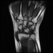
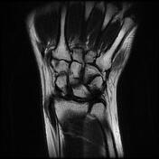
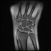
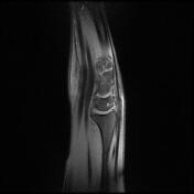
MRI reveals thickening with abnormal intrasubstance increased fluid signal of the first dorsal compartment tendons of the wrist (abductor pollicis longus and extensor pollicis brevis). There is increased fluid within the tendons sheath.
Abnormal intrasubstance increased fluid signal and focal thickening is present along with extensor carpi ulnar tendon (ECU) at the level of the wrist ulnar styloid and triangular fibrocartilage complex related to tendinosis.
TFCC central perforation is associated with fluid signal at distal radioulnar joint.
Small synovial cyst is seen at the volar aspect of the wrist radial side at the level of radioscaphoid joint.
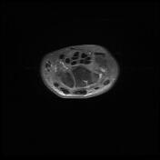
Intra osseous degenerative cyst within the lunate bone is seen.
Case Discussion
Clinical presentation and imaging findings are suggestive of De Quervain tenosynovitis.