
Presentation
Known hypertensive patient presents with altered mental status for last 3 days.
Patient Data
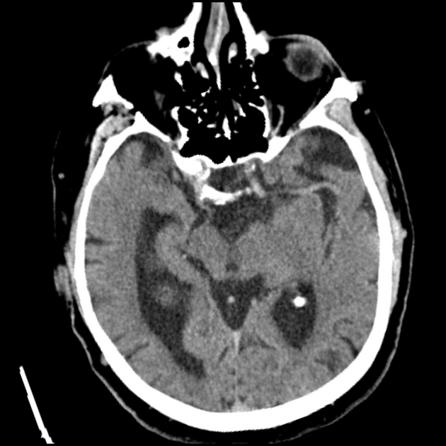
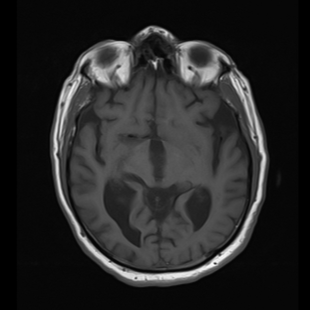
Age related brain parenchymal involution.
No intracranial hemorrhage.
No CT detectable territorial ischemic changes.


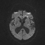
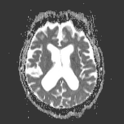
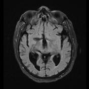
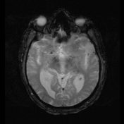
Multiple small T2 and FLAIR hyperintense foci oriented in a linear distribution in left frontal and parietal lobe periventricular white matter which are showing diffusion restriction, representing watershed acute ischemic infarcts in the border zone between left middle and anterior cerebral arteries.
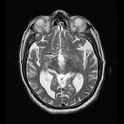
Note absence of normal flow signal in the left internal carotid artery below the base of skull presumably due to severe carotid stenosis.
Case Discussion
Watershed cerebral infarctions (border zone infarcts) occur at the border between cerebral vascular territories mainly in elderly.
Deep (internal) border zones infarct are mainly seen as ≥3 lesions, each ≥3 mm in diameter in a linear fashion parallel to the lateral ventricle in the centrum semiovale (Such in our case) or corona radiata, which sometimes become more confluent and band-like.
CT may fail in detection of early watershed infarcts, as in this case while diffusion weighted images are very helpul.


 Unable to process the form. Check for errors and try again.