
Presentation
This patient with a history of alcoholic liver disease was admitted to the ED with a clinical stroke.
Patient Data
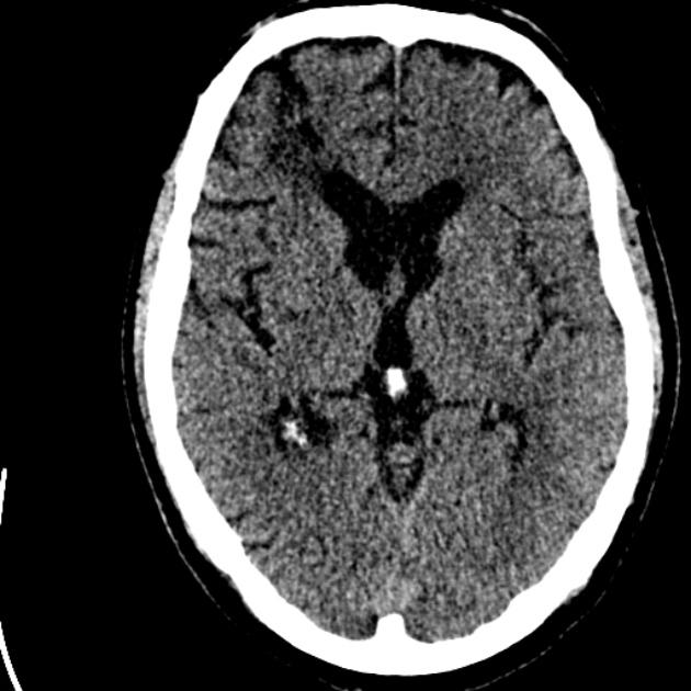
No evidence of acute hemorrhage or large cortical territorial infarction. There is some cerebral atrophy and an old right frontal lobe insult, along with some white matter changes typical for mild small vessel chronic ischemia.
The patient was commenced on aspirin for presumed stroke. While awaiting an MRI brain to confirm, the patient deteriorated with more pronounced signs of stroke. He then suffered a respiratory arrest.
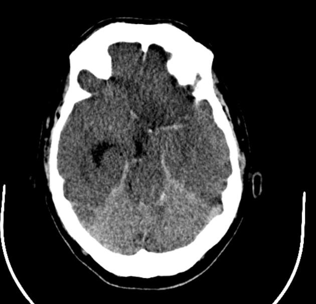
36 hours post admission CT.
There is acute hemorrhage in the posterior cranial fossa involving the pons and cerebellum, extending into the fourth ventricle.
Supratentorially, there is severe mass effect with midline shift to the right and sulcal effacement. Although there is obstructive hydrocephalus, the left lateral ventricle is completely compressed. There is hypoattenuation and tumefaction of the left MCA and ACA territories with subfalcine herniation.
The cerebellum appears of increased density against the hypoattenuating cerebrum.
Case Discussion
The appearances were considered incompatible with survival.
The exact sequence of events is unclear, but the respiratory arrest has likely resulted from the brainstem herniation, with associated Duret hemorrhage. ACA infarction is a recognized sequelae of subfalcine herniation causing ACA compression.


 Unable to process the form. Check for errors and try again.