
Presentation
Palpable lump in the left breast.
Patient Data

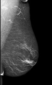
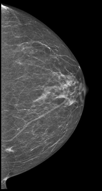
CC view of the left breast demonstrates a 10 mm asymmetry in the medial left breast posterior depth. No correlating abnormality on the MLO view.
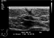
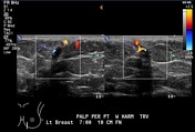

Sonographic evaluation of the palpable area, demonstrates a 10 mm irregular hypoechoic mass with vertical orientation, in the lower inner quadrant of the left breast, in the 7 o'clock position, 10 cm from the nipple. There is a track to the skin. There is internal vascularity on colour Doppler.
Case Discussion
This is a case of a 65-year-old female who presented for workup of a palpable mass in the left breast. Imaging features were suspicious for malignancy. The patient underwent a US-guided core biopsy with pathology consistent with desmoid fibromatosis of the breast. Immunohistochemical stain for Beta-catenin was strongly positive. The patient underwent surgical excision.
Desmoid fibromatosis of the breast is a rare and locally invasive benign tumour, resulting from abnormal proliferation of fibroblasts and myofibroblasts. Breast imaging findings are nonspecific and usually mimic malignancy. Standard treatment involves wide local excision.


 Unable to process the form. Check for errors and try again.