
Presentation
Head injury.
Patient Data
Age: Adult
Gender: Male
From the case:
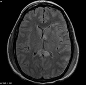
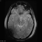
Diffuse axonal injury

Download
Info

Note the multiple sites of edema and hemorrhage, involving the brain stem and corpus callosum as well as subcortical white matter and left cerebral peduncle. High FLAIR signal is also seen in the dorsal midbrain. EVD insitu.
Case Discussion
Diffuse axonal injury can be subtle on CT but have devastating consequences for the patient. This is a case of grade III injury (involvement of brainstem) and carries a poor prognosis.


 Unable to process the form. Check for errors and try again.