
Presentation
Discoid, injured lateral meniscus confirmed during arthroscopy in the contralateral knee after trauma.
Patient Data
Age: 25 years
Gender: Male
From the case:
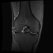
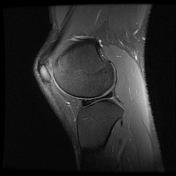
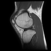
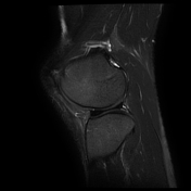
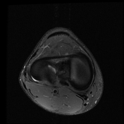
Discoid lateral meniscus

Download
Info

Thick, discoid, but intact, stable (meniscofemoral ligaments normal) lateral meniscus. >80% coverage indidcative of complete form.
Case Discussion
Discoid meniscus is often present bilaterally, the othwerwise asymptomatic right knee joint was imaged fot this reason.


 Unable to process the form. Check for errors and try again.