
Presentation
Knee pain, not specified.
Patient Data
Age: 65 years
Gender: Male


Download
Info
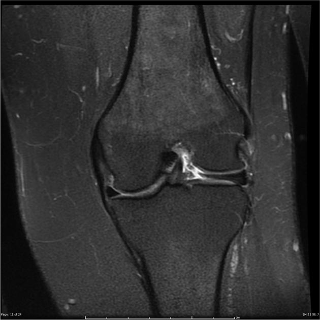
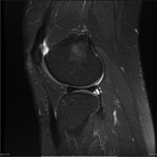
There is a discoid lateral meniscus without evidence of a lateral meniscal tear. Loss of articular cartilage of the weight-bearing portions of the femoral condyle and medial tibial plateau with a small region of focal full thickness loss in the central medial femoral condyle.
A 22 x 6 x 5 mm intra-articular body lies just posterior to the PCL. No evidence of a joint effusion. The ACL, PCL, MCL and LCL are intact. The posterolateral corner structures are intact. The patellar and quadriceps tendons are normal.


 Unable to process the form. Check for errors and try again.