
Presentation
Chronic bilateral knee pain.
Patient Data


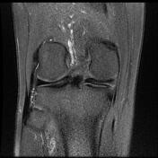
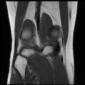

incomplete discoid medial meniscus with horizontal tear along with body and posterior horn
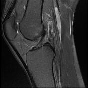
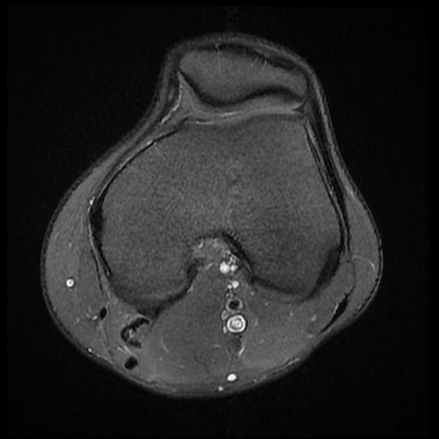
lateral meniscus with horizontal tear along with anterior horn, associated with parameniscal cyst measuring about 5 x 15 mm next to the anterior horn
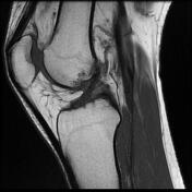
small popliteus tendon synovial cyst measuring about 5 x 20 mm

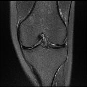

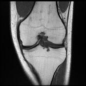
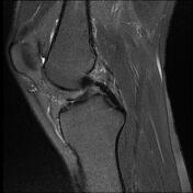
incomplete discoid medial meniscus with horizontal tear along with body and posterior horn
small popliteus tendon synovial cyst measuring about 10 x 20 mm
PCL ganglion cyst measuring about 5 x 10 mm
Case Discussion
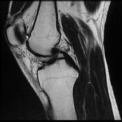
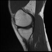
An unusual case of the bilateral incomplete discoid medial meniscus with a horizontal tear. Discoid meniscus is an anatomical variant that has a body that is too wide, usually affecting the lateral meniscus.


 Unable to process the form. Check for errors and try again.