Presentation
Knee pain.
Patient Data
Age: 20 years
Gender: Female

At the time the case was submitted for publication Hani M. Al Salam had no recorded disclosures.
View Hani M. Al Salam's current disclosuresKnee pain.


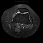
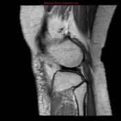
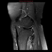
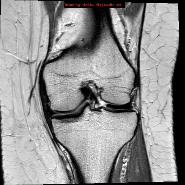
MRI knee demonstrates discoid lateral meniscus.
Public playlists
Unlisted playlists
This case is used in 24 unlisted playlists.
You can use Radiopaedia cases in a variety of ways to help you learn and teach.
Creating your own cases is easy.
ADVERTISEMENT: Supporters see fewer/no ads
Updating… Please wait.

 Unable to process the form. Check for errors and try again.
Thank you for updating your details.