
Presentation
Pain, swelling & clicking in right knee. History of trauma.
Patient Data
Age: 13 years
Gender: Female
From the case:
Discoid meniscus with tear


Download
Info
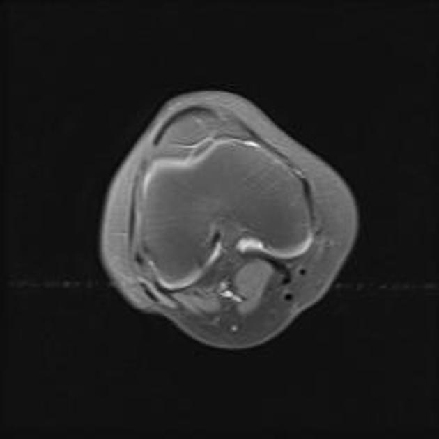
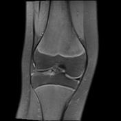
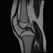
There is loss of normal semilunar shape of the lateral meniscus, which appears more than three consecutive sections on saggital images, represent discoid meniscus. It shows hyperintense signal on the T2W, PD FSAT images with flipped fragment on medial aspect, represents grade III meniscal signal
Case Discussion
- These findings represent discoid meniscus with Grade III meniscal signal.


 Unable to process the form. Check for errors and try again.