
Presentation
Post fall down wrist pain
Patient Data
Age: 35 years
Gender: Male
From the case:
Distal scaphoid fracture
Download
Info

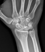
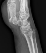
A plain radiograph revealed a lucency at the volar aspect of the distal scaphoid bone with suspected underlying fracture best seen on the lateral view
From the case:
Distal scaphoid fracture

Download
Info

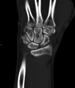
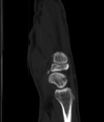
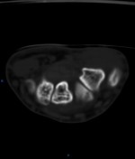
CT study confirms a distal scaphoid pole fracture with surrounding lucency possibly bone marrow contusion
From the case:
Distal scaphoid fracture

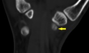
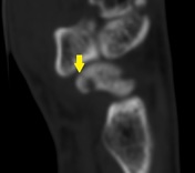

Download
Info
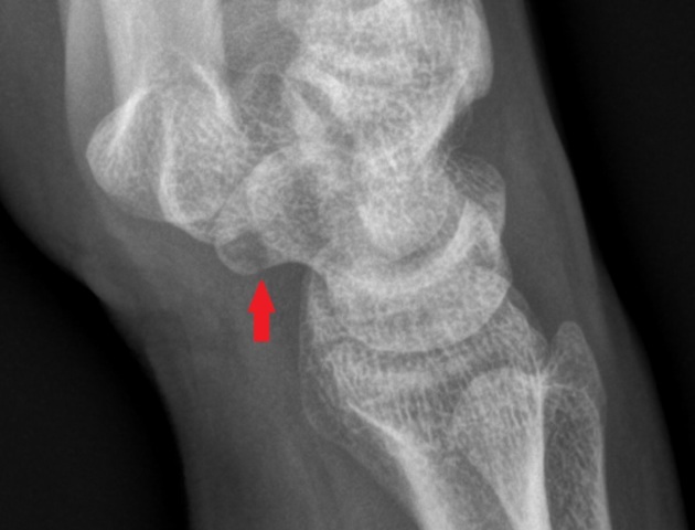
Annotated images highlight the findings in plain x-ray (the red arrow) and CT (the yellow arrows)
Case Discussion
I thought that this case should be shared as it is easy to be missed in the practice due to the less common occurrence. Scaphoid fractures commonly occur at the waist (70%-80%) while the distal pole represents 20% and the proximal pole 10%.


 Unable to process the form. Check for errors and try again.