
Presentation
Dyspnea, stridor and dysphagia.
Patient Data
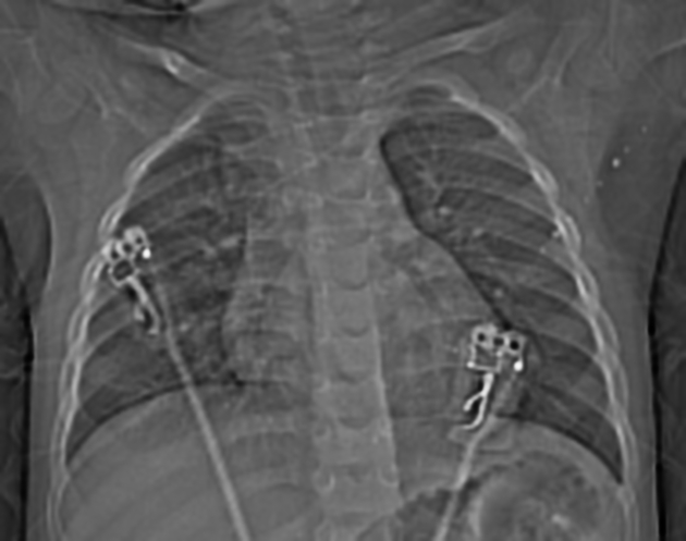
Narrowing of the air column in lower thoracic trachea with suggestion of a right sided arch.


Double aortic arch is seen with the ascending aorta is seen splitting into the right arch which is the larger and more superior and the left arch which is the smaller and at a lower level. The double aortic arch forms a vascular ring with compression of the proximal esophagus posteriorly as well as abutment and slight compression of the trachea. The double aortic arches unite posteriorly and form the descending aorta.
Two brachiocephalic arteries arise separately from the right and left arches with the four vessel sign.
No otherwise cardiac anomaly.
Case Discussion
Double aortic arch is the most common vascular ring due to persistence of the fourth aortic arches bilaterally. The ascending aorta is split into right and left aortic arches with the former is usually the dominant, higher and larger. The aortic arches join posteriorly to form the descending aorta.
The double aortic arches forms a complete symptomatic vascular ring that encircles the esophagus and trachea giving the symptoms of dyspnea and stridor (usually in the infancy and childhood) and dysphagia (more common in the adulthood).
Association of other congenital heart disease is rare.


 Unable to process the form. Check for errors and try again.