
Presentation
Routine screening mammogram left breast. No previous for comparison.
Patient Data

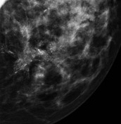
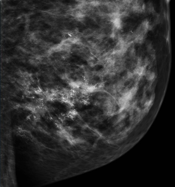
There is the "Mappa Mundi" of DCIS - the calcifications are "wild and crazy", there are X's, Y's and Z's visible haphazardly distributed on a segment of the breast. Look carefully at the distribution; you can see the calcifications in a ductal distribution as the DCIS grows out of blood supply and calcifies in response.
Case Discussion
This case shows all the hallmarks you would want to see in DCIS. In general, the wilder and crazier the microcalcifications, the higher the grade of DCIS.
Note superiorly how far the calcifications extend to the top of the image with "normal" breast in between. Mammography underestimates the extent of the disease; the degree and distribution of the calcifications are less than the disease extends into the breast. Just surgically removing the calcifications does not guarantee the disease has been completely removed. Hence the role of radiation in the therapy of DCIS.
In practice, ultrasound is useful. You can actually see these calcifications on ultrasound and biopsy them under ultrasound guidance.


 Unable to process the form. Check for errors and try again.