Presentation
Characterization of ovarian mass seen on US. History of pelvic pain.
Patient Data
Age: 30 years
Gender: Female


Download
Info

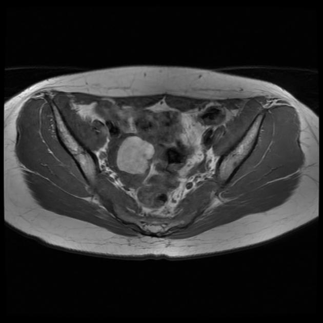
Selected series from a pelvic MRI study.
Right ovarian mass with uniformly low T2 and high T1 signal, low signal on DWI. Appearance in keeping with an endometrioma. Several normal ovarian follicles are noted adjacent to the endometrioma.
There is a further small focus of high T1 signal in the pouch of Douglas (best appreciated on the sag T1 fat sat sequence), suggestive of the presence of deep endometriosis.
Case Discussion
Typical MRI appearance of an endometrioma. There is also a likely focus of deep endometriosis.


 Unable to process the form. Check for errors and try again.