
Presentation
Progressively enlarged painful soft tissue bulge on the anterior inferior abdominal wall with a history of cesarian section fifteen years ago.
Patient Data


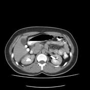
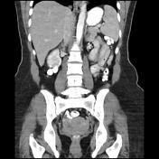
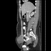
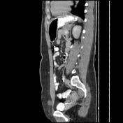
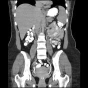
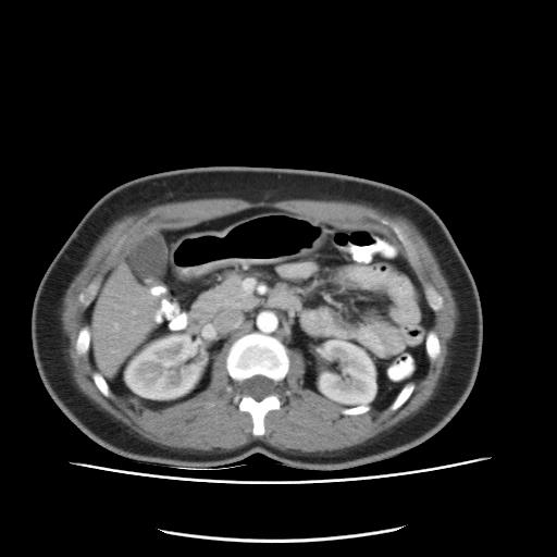
Large well-defined solid-cystic mass lesion axial width up to 90 x 80 mm and height up to 80 mm within anterior inferior abdominal wall rectus abdominis muscle partially extended within midline and contralateral rectus abdominis muscle is seen. The mass has a pronounced bulge within related subdermal fat and is minimally bulged within the related anterior pelvic cavity. Hypodense focus with size up to 25 x 18 mm in upper medial segment VIII of the right hepatic lobe with enhancement consistent with a hemangioma.
Case Discussion
The case illustrates the contrast-enhanced MDCT features of the pathology-proved abdominal wall cesarian section scar endometriosis. Wide surgical excision is the mainstay of treatment 1. This kind of endometriosis can be more common in cesarian sections with Pfannenstiel incision 2.


 Unable to process the form. Check for errors and try again.