
Presentation
Headache.
Patient Data


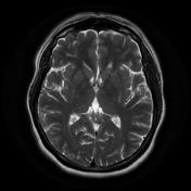
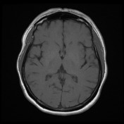
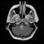
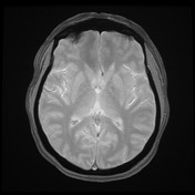

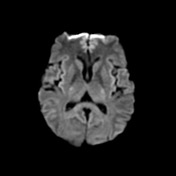
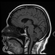
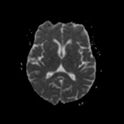
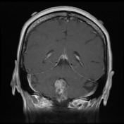
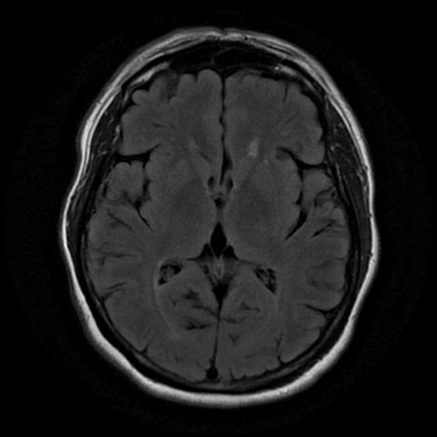
A lobulated heterogeneously enhancing tumor centered along the floor of the fourth ventricle demonstrates mild FLAIR hyperintensity with internal areas of FLAIR hypointensity/T2 hyperintensity, regions of mild T1 hyperintensity, and patchy susceptibility. Ao diffusion restriction. No surrounding edema. It traverses foramen Magendie into the cisterna magna. No extension through either of the foramina of Luschka. There is some mass effect on the tegmentum of the medulla, but is no distortion of the fourth ventricle and no hydrocephalus. No enhancing abnormality elsewhere.
Conclusion: Solitary heterogeneous posterior fossa tumor with cystic components and hemorrhage/calcifcation, protruding through the foramen of Magendie, most likely an ependymoma.
Case Discussion
The patient went on to have a resection.
Histology
MICROSCOPIC DESCRIPTION: Sections show a hypercellular tumor with prominent perivascular pseudorosettes. The tumor focally forms papillae and well formed ependymal canals. Tumor cells contain abundant pale eosinophilic to clear cytoplasm, oval nuclei with coarse granular chromatin and inconspicuous nucleoli. Focally tumor cells demonstrate mild to moderate nuclear atypia. There are up to 4 mitoses per 10 HPF identified. No necrosis or microvascular proliferation are seen.
Immunohistochemistry results show tumor cells stain:
- GFAP positive
- EMA positive (perinuclear dot staining)
- S100 negative
- Synaptophysin negative
- The Ki67 proliferation index is approximately 5%.
FINAL DIAGNOSIS: Ependymoma (WHO grade II).


 Unable to process the form. Check for errors and try again.