
Presentation
Left trigeminal neuralgia
Patient Data


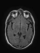
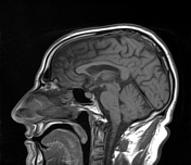
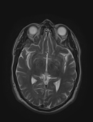
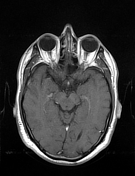
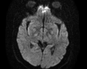

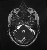
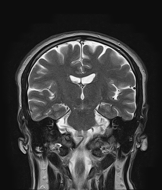
The MRI sequences demonstrate an extra-axial mass of irregular margins at the left cerebellopontine angle, extending into the ipsilateral internal auditory canal (ICA), encasing the facial/vestibulocochlear nerves, and compressing the cisternal portion of the ipsilateral trigeminal nerve. It is of low signal on T1, heterogeneous signal on FLAIR and high signal on T2 with no enhancement seen on the postcontrast sequence. It appears of high signal on DWI with low ADC (restricted diffusion).
Case Discussion
MRI features characteristic of a cerebellopontine angle epidermoid cyst compressing the trigeminal nerve.
DWI is important to differentiate epidermoid cyst from an arachnoid cyst ( the arachnoid cyst follows the CSF signal on all sequences with no restricted diffusion).


 Unable to process the form. Check for errors and try again.