
Presentation
Chronic flank pain
Patient Data

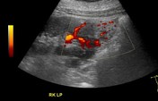
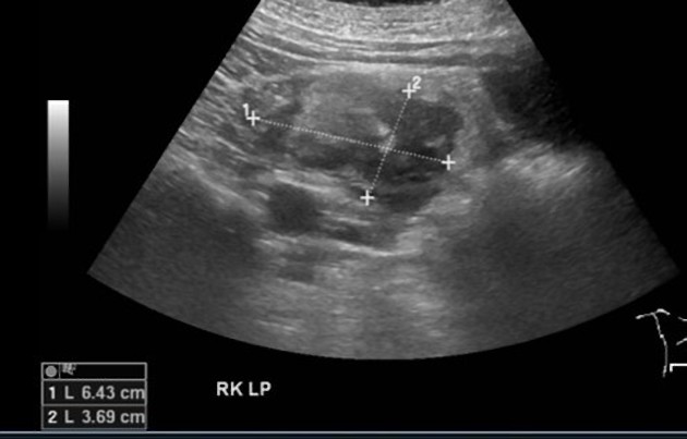
Ultrasound of the right kidney shows a large, relatively well defined heterogeneous, mainly hyperechoic lower pole cortical based renal mass with peripheral vascularity seen on color Doppler. A vessel seen extending from the renal cortex.


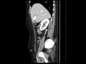

Axial reconstructions ( corticomedullary and nephrogenic phases) show a hypervascular exophytic mass arising from the lower pole of the right kidney, extending posteriorly into the perirenal compartment. There is a prominent draining vein into the IVC. No local infiltration or lymphadenopathy.
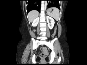
Coronal and sagittal reconstructions (nephrogenic phase) show the same mass with prominent draining vein at its posteroinferior aspect.
Pathology report
Sections showed tumor composed of three components, benign mesenchymal neoplasm composed of admixture of thick dysmorphic blood vessels, smooth muscle and small amount of adipose tissue. Cytological atypia and giant cells were seen in the smooth muscle. No increased mitosis or necrosis was identified. Epithelioid morphology was noted.
Impression:
Angiomyolipoma, likely epithelioid variant.
Case Discussion
Differential diagnoses
Epithelioid angiomyolipoma is a rare variant of angiomyolipoma; which can mimic renal cell carcinoma on imaging, both with less visible fat (microscopic). It is mainly a histological diagnosis, where biopsy might be indicated for differentiation.
-Classic angiomyolipoma : contains macroscopic fat; although can be still rarely seen in RCC.
-Oncocytoma: might vary in appearance and enhancement depending on its size. It might contain calcification or exhibit a non enhancing central stellate scar.
-Renal metastasis: can be suspected in case of known primary. It is more likely to be bilateral and multiple.


 Unable to process the form. Check for errors and try again.