
Presentation
Urinary obstruction of unknown cause.
Patient Data


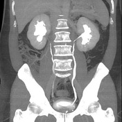
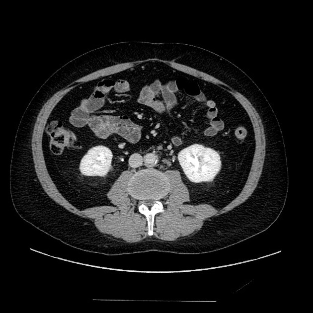
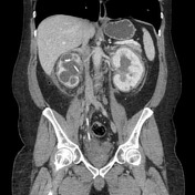
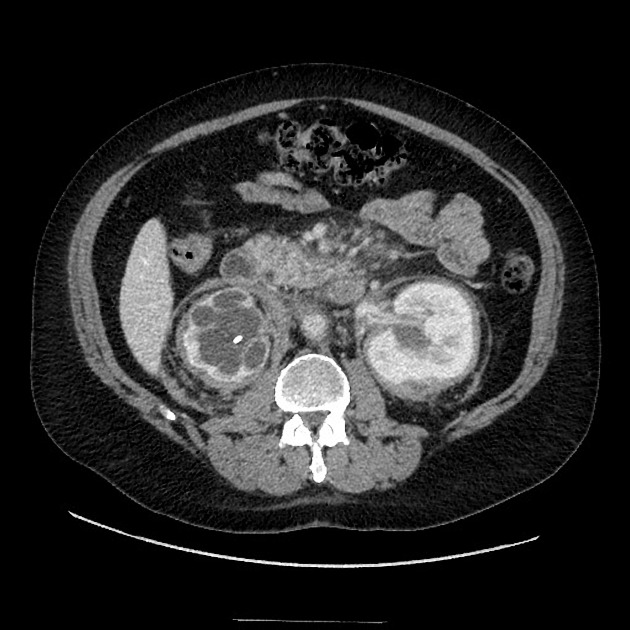
Ill-defined, strandy soft tissue in the renal hila and proximal ureters, resulting in mild bilateral hydronephrosis. This is fairly well seen on the coronal reformatted MIP images.
Soft tissue thickening surrounding portions of the left kidney.
Subtle short-segment circumferential thickening/enhancement of the mid-abdominal aorta.
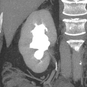
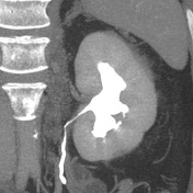
Similar appearance of mild bilateral hydronephrosis and soft tissue thickening/stranding in the renal hila/proximal ureters. Soft tissue thickening around the right kidney.
Similar short segment circumferential thickening surrounding the mid abdominal aorta.
Possibly early mild thickening surrounding the aortic arch.
Notable negatives: Normal adrenals and RCA.

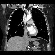

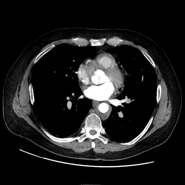
Increased circumferential soft tissue thickening or "coating" of the thoracic aorta and proximal great vessels.
Soft tissue surrounding the RCA and filling the right atrioventricular groove.
Partially imaged infiltration of the adrenals.

Soft tissue infiltration of the adrenals, right greater than left.
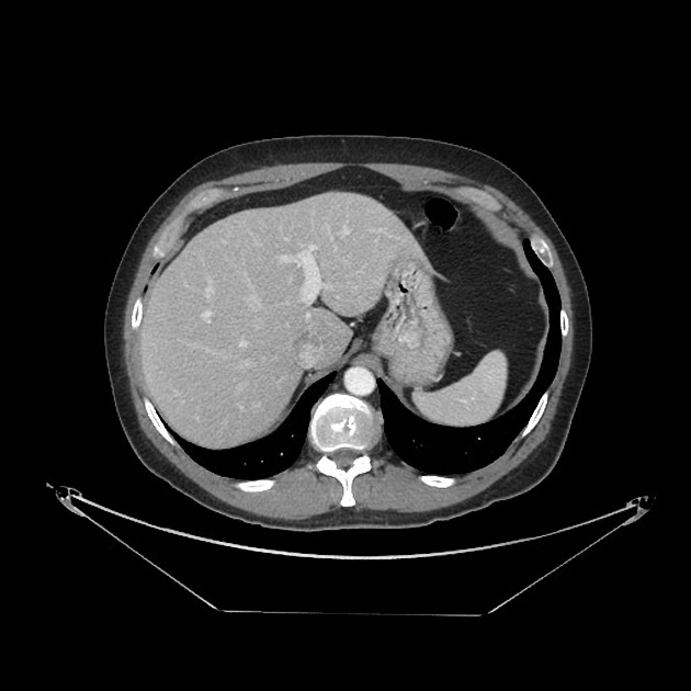
Thickening of the gallbladder and cystic duct.
Coating of the abdominal aorta and proximal common iliac.
Narrowing of the IVC with soft tissue infiltration.
Severely hydronephrotic right kidney with stent.
Mild hydronephrosis left kidney. Perinephric and perihilar soft tissue infiltration ("hairy kidney").
Case Discussion
Rare case showing a relatively gradual progression of findings typical for Erdheim-Chester disease. The patient presented with renal obstruction due to ill-defined, infiltrative soft tissue in the renal hila and proximal ureters.
Over the course of 9 years, the patient developed several more manifestations of the disease:
- increasing renal involvement, resulting in obstruction of the right kidney and typical "hairy kidney" appearance of the left
- adrenal infiltration
- biliary involvement with gallbladder and cystic duct thickening
- coating of the proximal great vessels and thoracic and abdominal aorta
- involvement and narrowing of the IVC
- infiltration of the RCA and right AV groove
Please see companion case with many overlapping imaging features.


 Unable to process the form. Check for errors and try again.