
Presentation
Bilateral hand pain and swelling.
Patient Data

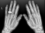
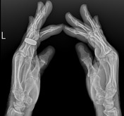
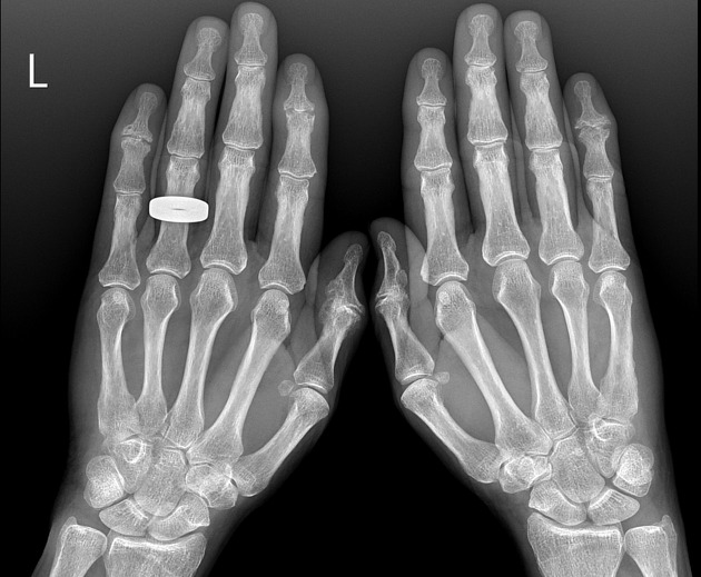
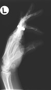
Joint space narrowing and marginal osteophyte narrowing of the distal interphalangeal joint row including the thumb interphalangeal joint. Central erosions are seen at the left 2nd and 5th, and right 5th. A "gull-wing" appearance is seen, most apparent of the right 5th distal interphalangeal joint on the ball-catcher's projection.
The proximal hand joints and carpus are spared. Normal alignment. No soft tissue calcification.
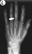
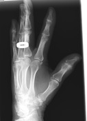

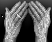
Distal interphalangeal row joint space narrowing and marginal osteophytes. Small central erosions are seen at the fifth distal interphalangeal joint.
Case Discussion
Even though the demographics are atypical, these changes are typical for erosive osteoarthritis, and the central erosions are especially convincing as they have increased in size and number between the two exams.


 Unable to process the form. Check for errors and try again.