
Presentation
Right proptosis.
Patient Data
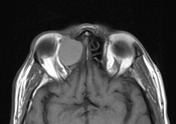

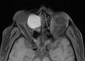
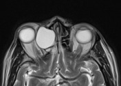

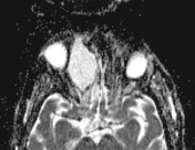

Right anterior ethmoidal mucocele is noted, measuring 26 x 28 mm in axial dimensions and showing expansile growth with homogeneous T1 and T2 hyperintensity. No diffusion restrictions.
This is associated with expansile thinning of the surrounding bones, mounting to complete dehiscence of the anterior part of the lamina papyraceae. There is extrinsic compression of the medial extraconal space of the right orbit, stretching the medial rectus and causing proptosis. Also noted partial focal dehiscence of the lateral lamella and fovea ethmoidalis.
The right frontal sinus is opacified by retentional phenomena.
Small retention cysts/mucosal polyps in both maxillary antra.
Marked rightwards nasal septum deviation with bony spur.
The remaining sinonasal cavities are otherwise unremarkable.
Case Discussion
Ethmoid sinus is the second most common site for paranasal sinus mucocele after frontal sinus. It is due to obstruction of the sinus ostium by secretions, inflammation or mass lesion. Sinus secretions accumulate over a long period with resultant sinus expansion, bone remodeling and thinning with local mass effect.
Ethmoid mucocele can cause proptosis (as in this case) or orbital apex compression. Superadded infection can lead to extension of the inflammatory process to nearby anatomical structures with formation of subcutaneous abscess (Pott's puffy tumor), epidural abscess or subperiosteal orbital abscess.


 Unable to process the form. Check for errors and try again.