
Presentation
Fall from height.
Patient Data
Age: 85 years
Gender: Female
From the case:
Extensive subgaleal hematoma


Download
Info
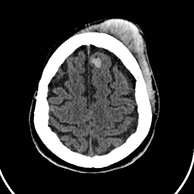
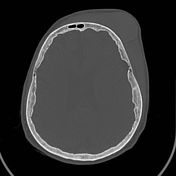
Extensive left frontotemporal subgaleal hematoma (hyperattenuating content) associated with soft tissues edema in adjacent face, as well a small intraparenchymal subcortical contusion in the left frontal lobe.
No associated skull fractures.
Download
Info
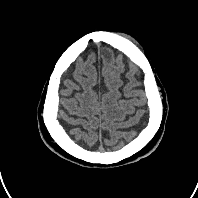
CT scan done 15 days later showed an impressive reduction/absorption of the subgaleal hematoma. The intraparenchymal hemorrhage (contusion) was resolved, remaining a small focus of hypoattenuation.
Case Discussion
A subgaleal hematoma describes bleeding in the potential space between the periosteum and the galea aponeurosis.
A cerebral hemorrhagic contusion is a type of intracerebral hemorrhage and is common in the setting of significant head injury.


 Unable to process the form. Check for errors and try again.