Presentation
Left external auditory canal mass on physical examination
Patient Data
Age: 20 years
Gender: Male
From the case:
External auditory canal osteoma


Download
Info

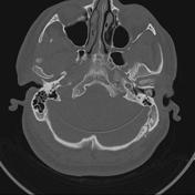
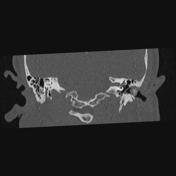
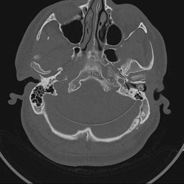
Pedunculated bony mass arising from the anterior wall of the left external auditory canal and almost obliterating the canal.
Case Discussion
The findings are consistent with left external auditory canal osteoma.
Osteomas of the external auditory canal are considered clinically to be discrete, pedunculated bone lesions arising along the tympano-squamous suture while exostoses of external auditory canal are broad based elevations of bone usually multiple and bilaterally symmetric, involving the tympanic bone.


 Unable to process the form. Check for errors and try again.